


The glaucoma consultation is an in-depth consultation that includes an eye examination as well as specialised tests to monitor and assess structures of the eye relating to glaucoma. It is designed for people who:
- have been defined as at-risk (for example, you have borderline eye pressure).
- Over 40 year olds who have a mother, father, brother or sister with glaucoma.
- Have been diagnosed with glaucoma and want additional monitoring to ensure the condition is well-controlled with the medical therapies prescribed.

Who is the Glaucoma Consultation for?
The glaucoma consultation is an in-depth consultation that includes an eye examination as well as specialised tests to monitor and assess structures of the eye relating to glaucoma. It is designed for people who:
- have been defined as at-risk (for example, you have borderline eye pressure).
- Over 40 year olds who have a mother, father, brother or sister with glaucoma.
- Have been diagnosed with glaucoma and want additional monitoring to ensure the condition is well-controlled with the medical therapies prescribed.
Why Have the Glaucoma Consultation?
The standard eye examination is a screening assessment for general eye conditions and a sight test. It will identify that you are at risk based on a series of tests that every optometrist will be able to perform. However, to be able to diagnose glaucoma early more sophisticated diagnostic tests are required. These assessments are not available in every practice as the technology is expensive.
The standard eye examination basically measures eye pressure (most often with a puff of air, which is not the gold standard) and a basic assessment of the optic nerve and sometimes a visual field test. All three of these tests are not the best way to diagnose glaucoma but together are the minimum level required to form an adequate screening examination (together with factors like family history).
What is the Gold Standard of Detection & Monitoring?
The gold standard of glaucoma assessment involves performing the following tests:
- Dilated binocular examination of the optic nerves
- 3D OCT scan with glaucoma progression analysis
- Assessment of corneal thickness
- Measuring eye pressure with Goldmann tonometry
- Gonioscopy or anterior segment OCT
- Full threshold visual fields
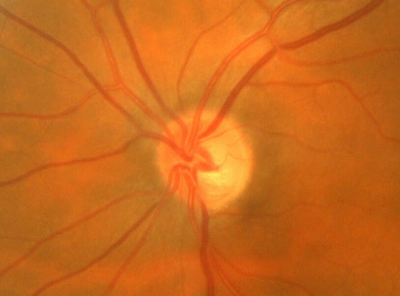
1. Dilation & Examination of the Optic Nerves
As we grow older the pupils get smaller. It is much easier for the Optometrist get a clear view of the optic nerve after instilling drops that temporarily open up the pupil, so that a clear view of the optic nerve can be made.
A photograph of the optic nerve is captured for future reference.
2. 3D OCT Scans
3D OCT or Optical Coherence Tomography is the most sophisticated way to evaluate the optic nerve. However, the area of glaucoma monitoring and detection with OCT is constantly changing with newer better approaches through innovations in OCT computer software. We’ve invested in our 3rd OCT with the latest glaucoma analysis modules.
We also keep up-to-date with the latest articles in peer-reviewed journals to ensure offering the best level of care to our patients.
At the present time we use a variety of OCT scans to help diagnosis of glaucoma. We will perform a wide field scan and also a macula scan. If it is your first appointment we will compare the results with a database of normal eyes in order to benchmark your results. We will also compare the results with other tests such as more detailed 24-2 and 10-2 visual field tests.

3. Corneal Thickness
Measuring the thickness of the cornea is very important. A large study showed a drammatic association between thinner corneal measurements and the development of glaucoma. At the 5-year time point in the study, 36% of individuals who at presentation had eye pressure higher than 25.75 mm Hg (average 27.9) and a corneal thickness of less than 555 µm (average 532) developed glaucoma, while only 6% of those with the same eye pressure but with corneas thicker than 588 µm (average 613.4) developed glaucoma.
4. Goldmann Tonometry
Accurate measurement of eye pressure is important in glaucoma diagnosis as eye pressure is associated with glaucoma development. That said, the relationship is not clear-cut. Some people with mildly raised pressures develop glaucoma and others do not.
Eye pressures are normally measured with an air-puff device. However, when the eye pressure is in the early 20’s these devices often over-estimate eye pressure. We therefore always used Goldmann tonometry which is the same device used in eye hospitals. Most people find it is more comfortable than the air-puff. A local anaesthetic is used to numb the eyes for a short time whilst eye pressure is measured. It only takes around a minute to take eye pressures.
5. Gonioscopy & Anterior Segment OCT
Examination of the drainage system is important for several reasons. Firstly, there isn’t just one type of glaucoma. The most common is primary open angle glaucoma. However, angle closure glaucoma can only be properly diagnosed by looking at the drainage system to see how open it is for the fluid in the eye to leave without obstruction. There can also be other, more rare, causes of obstruction which need to be detected.
The best way to look at the angle is with Gonioscopy and Anterior segment OCT. Gonioscopy involves placing a special contact lens on the eye. The lens has mirrors which allows our Optometrists to see the drainage system.
Anterior segment OCT is a new way of looking at the angle. It allows us to see angle and also behind the iris.
6. Full Threshold Visual Fields
Damage to the optic nerve ultimately causes a reduction in the field of vision. The visual field test in a test that evaluates the function of the optic nerve. It provides us with information relating to how you field of vision is affected by glaucoma.
We perform full threshold fields in the 2 key areas where glaucoma causes field loss. These are the 24 degree and 10 degree tests. Full threshold means we assess the visual field at the threshold of the ability of the retina to detect the test target 50% of the time. There are variety of outputs from the fields test and these need to be carefully interpretted in order to assess the result.
We use the field test to monitor progression or changes and also to compare the location of an field loss to structural damage detected using the OCT scan.
Vision Plan Elite
Vision Plan Elite was designed specifically to cater for patients looking for full, frequent monitoring and assessments of their eyes using our very latest technology and comprehensive examination protocols. You’ll need to come into the practice to sign-up for Vision Plan Elite by filling in a Direct Debit form.
Vision Plan Elite £15 / month
(On-going specialist examinations)
Terms & Conditions
- Vision Plan Elite covers all specialty consultations and initial treatment consultations.
- A single monthly fee covers any additional assessments with the access to all our technology.
- Ideally suited to patients in need of regular monitoring of eye conditions like glaucoma or macular degeneration.
- 20% off all frames and lenses.
- Priority appointments.
- Free adjustments and repairs on glasses.
- Exclusive invitations to open days.
- Vision Plan Elite is a 18 month rolling contract.
Book One-Off Consultation
Vision Plan Elite
Vision Plan Elite was designed specifically to cater for patients looking for full, frequent monitoring and assessments of their eyes using our very latest technology and comprehensive examination protocols. You’ll need to come into the practice to sign-up for Vision Plan Elite by filling in a Direct Debit form.
Vision Plan Elite £15 / month
(On-going specialist examinations)
Terms & Conditions
- Vision Plan Elite covers all specialty consultations and initial treatment consultations.
- A single monthly fee covers any additional assessments with the access to all our technology.
- Ideally suited to patients in need of regular monitoring of eye conditions like glaucoma or macular degeneration.
- 20% off all frames and lenses.
- Priority appointments.
- Free adjustments and repairs on glasses.
- Exclusive invitations to open days.
- Vision Plan Elite is a 18 month rolling contract.